Halal Empty Capsule,Halal Empty Gelatin Capsules,Empty Capsule Shell With Halal,Halal Shell Empty Gelatin Capsules Ningbo Jiangnan Capsule Co., Ltd. , https://www.jncapsule.com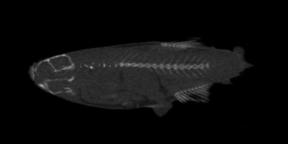

Introduction of zebrafish micro-CT experiment
Introduction of zebrafish micro-CT experiment
As a traditional vertebrate model, zebrafish has been widely used in the study of human diseases and embryonic development. The whole zebrafish gene is completely clear and has 85% homology with the human genome, which means experiments in zebrafish. Many of the results apply to humans. Zebrafish has higher reproductive rate and growth rate than other commonly used animals, and its embryonic development process is carried out in vitro. The researchers directly observe the zebrafish fertilized eggs through the microscope to the blastocyst stage and then develop into small fish. The whole process. This feature is of great significance for research on embryo development and research in genetic engineering, and is easy to perform gene knockout and knock-in. In addition, compared with commonly used model animals such as mice and rats, zebrafish have lower breeding costs, stronger reproductive ability, and easier access to a large number of individuals, facilitating the advantages of large sample size studies. Therefore, zebrafish has been widely used as a model organism in the field of life science research.
Micro computed tomography (micro computed tomography), also known as micro CT, small animal CT, is a non-destructive 3D imaging technique that clearly understands the internal microscopy of a sample without destroying the sample. structure. The biggest difference between it and ordinary clinical CT is that it has a very high resolution and can reach the micrometer (μm) level, so it has a good "microscopic" effect. Small animal CT can be used in the fields of medicine, pharmacy, biology, archaeology, materials, electronics, geology and so on. At present, many foreign researchers use this technology to conduct research in the field of orthopedics. Through this technology, it is possible to obtain a plurality of commonly used and important parameter data of the bone, and the scanned image can visually observe the change of the bone, including the morphological structure change of the trabecular bone.
This experiment was an attempt to observe the skeletal structure of zebrafish using micro-CT. The use of zebrafish for orthopedic research is now common, because zebrafish facilitate gene knockout and knock-in during embryonic period, which is beneficial for us to study the effects of a gene on animal bones. Micro-CT has high resolution and can analyze bone parameters of bone structure, which is an advanced research technology in orthopedic research. If the zebrafish after mutation can be observed by micro-CT, it will open a new page for the field of orthopedic research!
The instrument used in this experiment is the ZKKS-MCT-Sharp-V micro-CT of Zhongke Yusheng. The scanning parameters are voltage 50Kvp, power 50W, 4 frames superimposed, and the field of view is 4 cm×4 cm. The experimental material was selected for 90-day zebrafish and scanned after fixation.
experiment procedure:
The zebrafish is first fixed by a mold, scanned, and reconstructed after the scan, and then local reconstruction is performed according to the region of interest, and bone analysis is performed after local reconstruction.
The experimental results are as follows:
Figure 1 Two-dimensional longitudinal section of zebrafish micro-CT scan
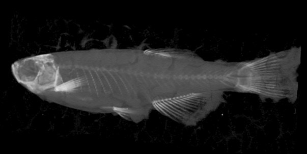
Figure 2: Two-dimensional slice of zebrafish micro-CT scan
Figure 3: Three-dimensional longitudinal section of zebrafish micro-CT scan
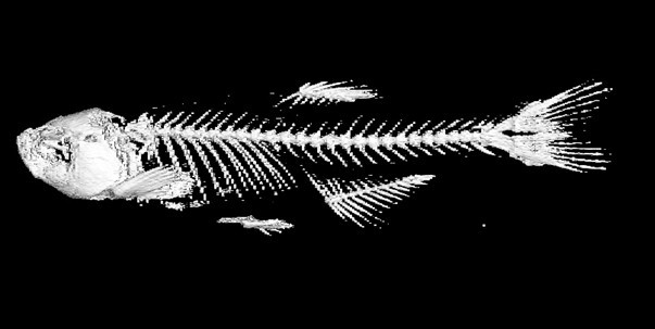
Figure 4: Zebrafish bone parameters analysis bone 3D map
Figure 1 and Figure 2 are two-dimensional slice diagrams obtained after partial reconstruction after scanning. Figure 1 is a vertical slice diagram cut along the Y-axis, and Figure 2 is a slice diagram cut along the Z-axis. As can be seen from the figure, micro-CT can clearly scan the skull, vertebrae, ribs and fin bones. Figure 3 shows the three-dimensional image after partial reconstruction. You can see the translucent muscle-wrapped bone structure. This picture shows the skeleton structure of the zebrafish more clearly and completely. All the above three pictures show that the microscopic CT can be used to visually observe the morphological observation of the zebrafish bone structure. The fourth picture is a picture after bone analysis. In this picture, the muscles and other tissues are removed, leaving only the bone structure, and it is possible to study the bones at a glance.
In addition to these intuitive images, a series of bone parameters can be obtained through bone analysis, such as the parameters BMC, TMC, BMD, TMD, Tb.N, Tb.Sp that are often of interest in the study of osteosclerosis and osteoporosis. , Tb.Th, Tb.Pf, etc. These data are useful for comparison to obtain quantitative indicators.
In summary, this attempt proves that micro-CT has important value in the study of zebrafish bones, and its advanced research methods will definitely bring great convenience to orthopedic research.